Skip to main content
search
contact@ardea.com
020 8318 1401
Explore
Inspiration
About Us
Contact Us
Account
Lightboxes
Print Store
Sign In
Home
>
Contributors
>
DSP
David Spears
240 Items
<
1
2
3
>
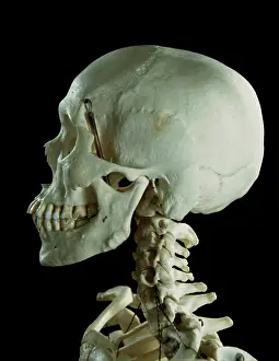
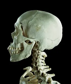
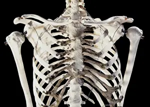
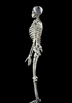
Human Skeleton body structure
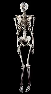
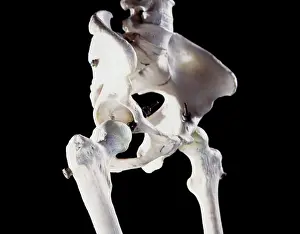
Human Skeleton body structure
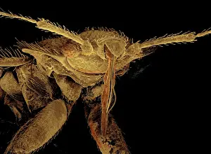
Bedbug
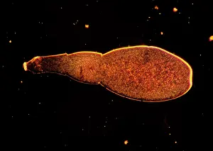
Light Micrograph: Tapeworm
Scanning Electron Micrograph (SEM): Leather or Hide Beet
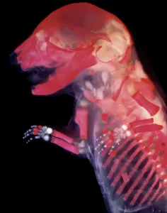
Mouse Embryo - red dye to show bones
Eye detail of a rat embryo at 13.2 days after fertilisation
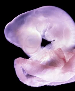
Rat Embryo 11.5 days after fertilisation
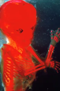
Human Foetus 12 weeks - red dye to show bones
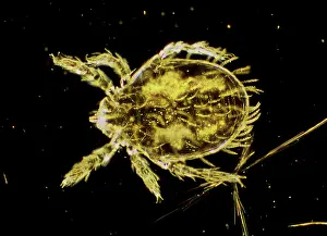
Dark Field Light Micrograph: Scrub Typhus Mite Chigger
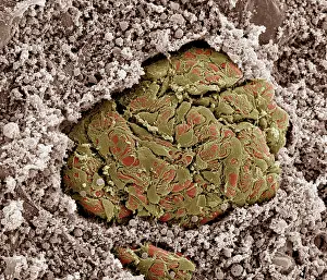
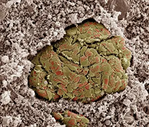
Scanning electron micrograph (SEM) of Kidney glomerulus
Scanning Electron Micrograph (SEM): Dinoflagellate
Scanning Electron Micrograph (SEM)
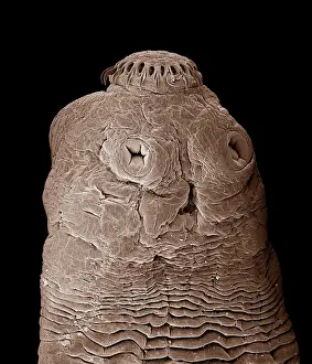
Tapeworm
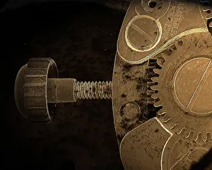
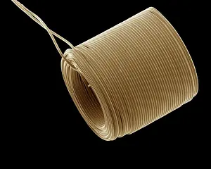
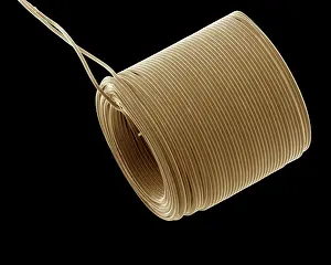
Scanning Electron Micrograph (SEM); Watch Winder
Scanning Electron Micrograph (SEM): Tardigrade or
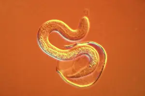
Light Micrograph using Nomarski Technique: Intestinal
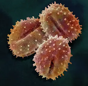
Scanning Electron Micrograph (SEM); Pollen of Mistletoe
Dark Field Light Micrograph (LM): Human Pinworm
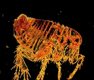
Dark Field Light Micrograph: Human Flea - male
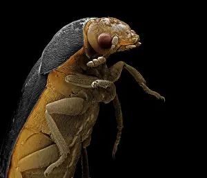
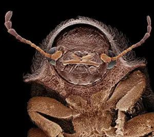
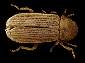
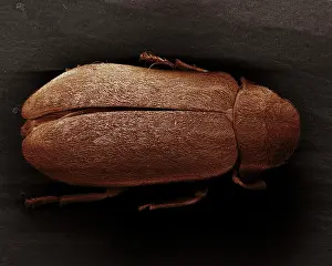
Scanning Electron Micrograph (SEM): Deathwatch Beetle
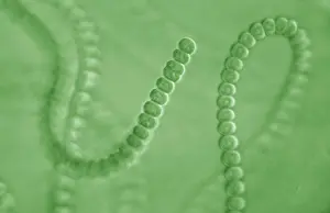
Light Micrograph: Cyanobacterium
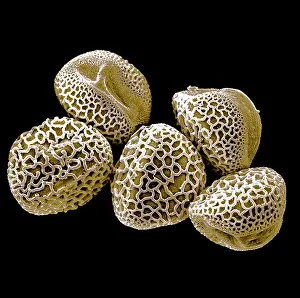
Scanning Electron micrograph (SEM): Lily Pollen grains
Scanning Electron Micrograph (SEM): a corrosion
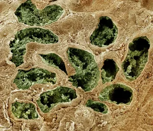
Scanning Electron Micrograph (SEM): Human testis
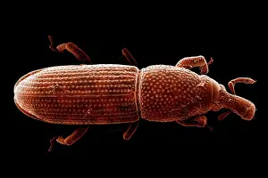
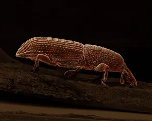
Scanning Electron Micrograph (SEM): Wood-boring Weevil
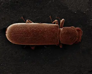
Scanning Electron Micrograph (SEM): Powder Post Beetle
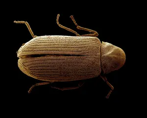
Scanning Electron Micrograph (SEM): Common Furniture
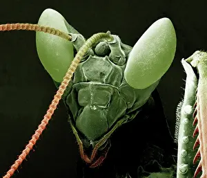
Scanning Electron Micrograph (SEM): Praying Mantis
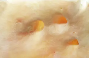
Atherosclerotic Aorta Vessel
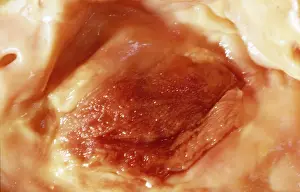
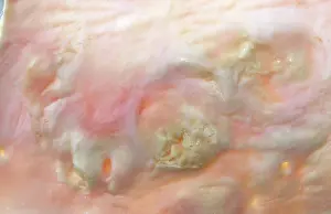
Atherosclerotic Plaque and Obstructed Lumen of Human Aorta
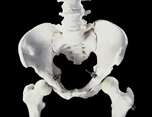
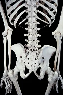
Human Skeleton
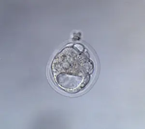
Cavitated Mouse Blastocyst
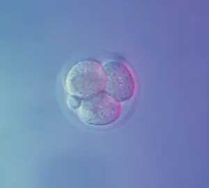
Mouse Embryo at Four Cell Stage & a Polar Body
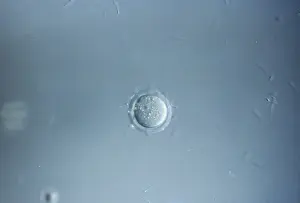
Mouse Ovum being fertilized
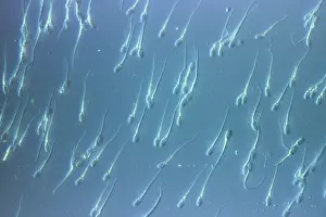
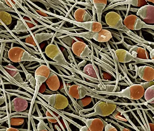
Human Sperm
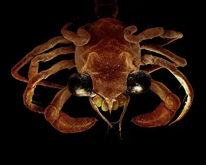
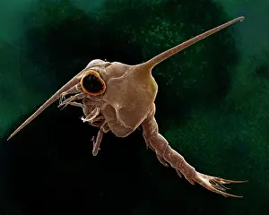
Scanning Electron Micrograph (SEM): Crab megalops
Scanning Electron Micrograph (SEM): Crab megalops
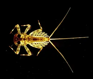
Light Micrograph Flattened Mayfly Nymph
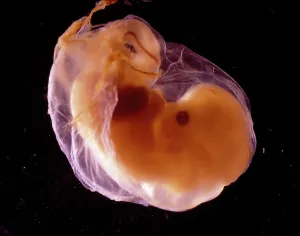
Human Embryo
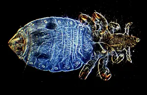
Dark Field Light Micrograph Human Head Louse
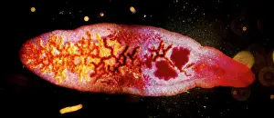
Dark Field Light Micrograph: Lancet Liver Fluke
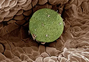
Scanning Electron Micrograph (SEM); Human Ovum in
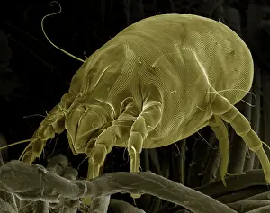
Scanning Electron Micrograph (SEM) Dust Mite
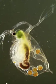
Light Micrograph: Water Flea
Scanning Electron Micrograph (SEM): Crab larvae
Scanning Electron Micrograph (SEM): American Cockroach
Scanning Electron Micrograph (SEM); Catheter
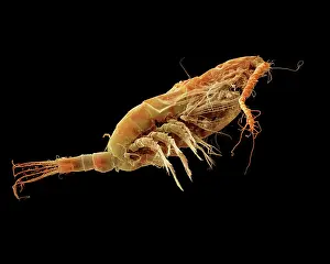
Scanning Electron Micrograph (SEM): Calanus finmarchicus
Macro Photograph
Scanning Electron Micrograph (SEM); Catheter
Scanning Electron Micrograph (SEM) showing fruiting
Scanning Electron Micrograph (SEM): Fusarium
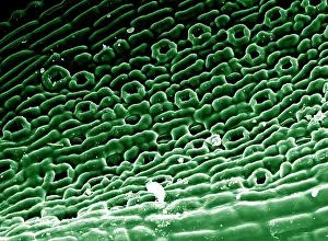
Scanning Electron micrograph (SEM)showing stomata
Scanning Electron micrograph (SEM): Grass Pollen grains
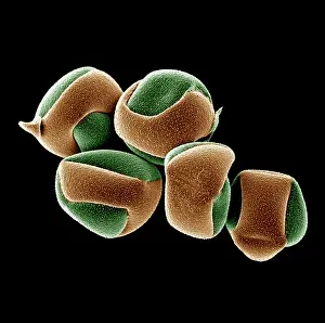
Scanning Electron micrograph (SEM): Cactus Pollen grains
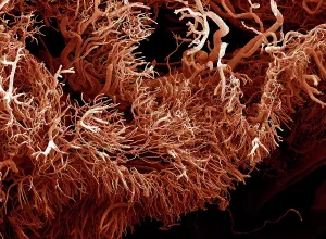
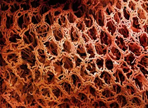
Scanning Electron Micrograph (SEM) of a resin cast
Scanning Electron Micrograph (SEM): Human Retina
Scanning electron micrograph (SEM) of Kidney glomerulus
Scanning Electron Micrograph (SEM): Human sperm
Scanning Electron Micrograph (SEM): Early human
Scanning Electron Micrograph (SEM): Human skin section
Scanning Electron Micrograph (SEM): Follicle Mite
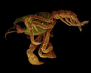
Scanning Electron Micrograph (SEM): False Scorpion
Scanning Electron Micrograph (SEM): Death Watch Beetle
Scanning Electron Micrograph (SEM): Powderpost Beetle
Scanning Electron Micrograph (SEM): Wood-boring Weevil
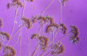
Light Micrograph showing fruiting bodies (conidiophores)
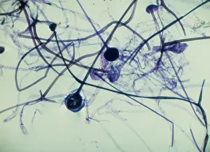
Light Micrograph (LM): Rhizopus sporangia
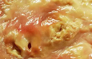
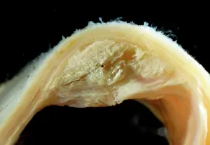
Atherosclerotic Plaque and Obstructed Lumen of Human Aorta
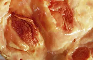
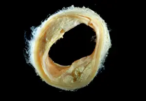
Atherosclerotic Plaque and Obstructed Lumen of Human Aorta
Atherosclerotic Plaque and Obstructed Lumen of Human Aorta
Atherosclerotic Plaque and Obstructed Lumen of Human Aorta
Atherosclerotic Plaque and Obstructed Lumen of Human Aorta
Atherosclerotic Plaque and Obstructed Lumen of Human Aorta
Atherosclerotic Plaque and Obstructed Lumen of Human Aorta
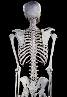
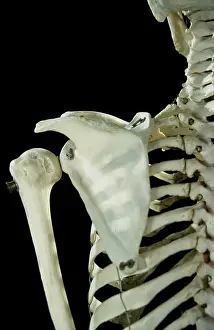
Human Skeleton body structure
Human Skeleton
Human Skeleton
Human Skeleton
Human Skeleton
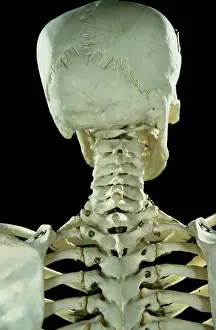
Human Skeleton body structure
Human Skeleton
Human Skeleton
<
1
2
3
>
© Copyright Ardea Picture Library - All Rights Reserved 2026
Powered by
Media Storehouse
®