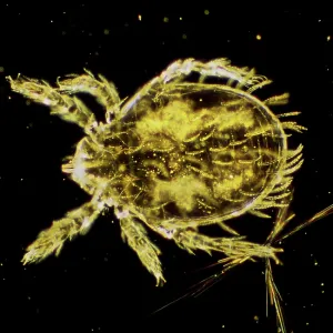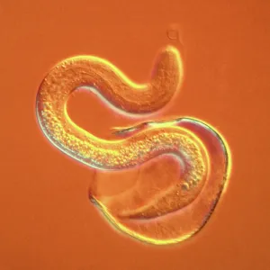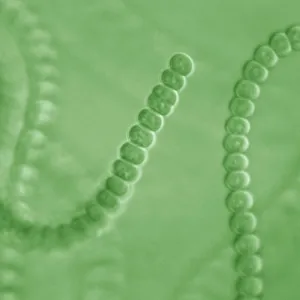
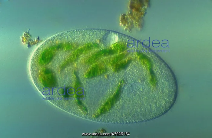


 : Light Micrograph: Ciliate


Light Micrograph: Ciliate


Picture No
11675491
Media ID
43026154
Size
6544px x 4272px (3.7MB)
Credit
© www.ardea.com / Ardea / David Spears~~
Category
NA
Ardea Picture Library
Picture No
11675491
Contributor Ref
DSP-141

Pick your License

Please supply intended use


Ciliate Ciliates Ciliophora Eukaryotic Micro Micrograph Micrographs Micrography Microorganisms Microscopic Plankton Protist Protists


Features In These Collections

> Contributors > DSP
Gallery Wall Inspiration

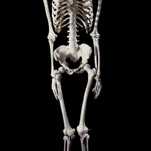
Human Skeleton body structure
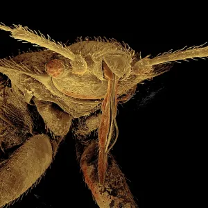
Bedbug
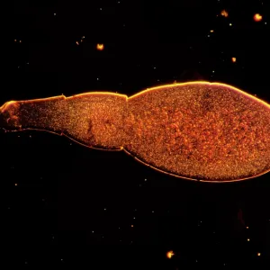
Light Micrograph: Tapeworm
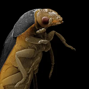
Scanning Electron Micrograph (SEM): Leather or Hide Beet
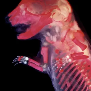
Mouse Embryo - red dye to show bones
Eye detail of a rat embryo at 13.2 days after fertilisation
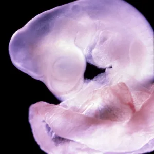
Rat Embryo 11.5 days after fertilisation
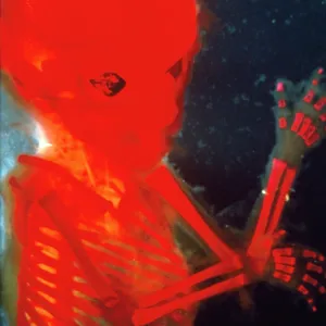
Human Foetus 12 weeks - red dye to show bones
Dark Field Light Micrograph: Scrub Typhus Mite Chigger
Scanning electron micrograph (SEM) of Kidney glomerulus
Scanning Electron Micrograph (SEM): Dinoflagellate
Scanning Electron Micrograph (SEM)
Tapeworm
Scanning Electron Micrograph (SEM); Watch Winder
Scanning Electron Micrograph (SEM): Tardigrade or
Light Micrograph using Nomarski Technique: Intestinal
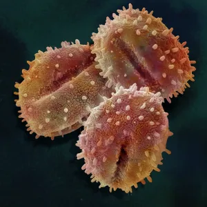
Scanning Electron Micrograph (SEM); Pollen of Mistletoe
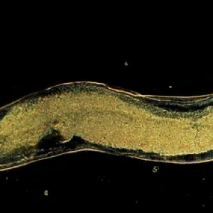
Dark Field Light Micrograph (LM): Human Pinworm
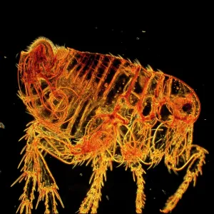
Dark Field Light Micrograph: Human Flea - male
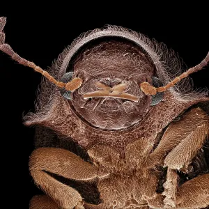
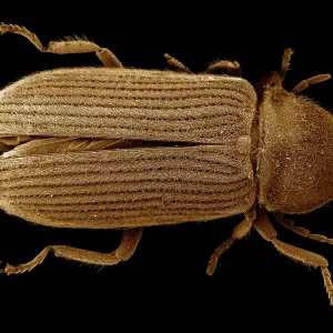
Scanning Electron Micrograph (SEM): Deathwatch Beetle
Light Micrograph: Cyanobacterium
Scanning Electron micrograph (SEM): Lily Pollen grains
Scanning Electron Micrograph (SEM): a corrosion
Scanning Electron Micrograph (SEM): Human testis
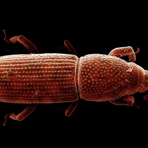
Scanning Electron Micrograph (SEM): Wood-boring Weevil
Scanning Electron Micrograph (SEM): Powder Post Beetle


© Copyright Ardea Picture Library - All Rights Reserved 2026
Powered by Media Storehouse®
We accept all major payment options so you can pay for the things you need in the way that is most convenient for you
Facebook     Twitter